Upper Thigh Cross Sectional Anatomy : Muscles Of The Posterior Thigh Hamstrings Damage Teachmeanatomy : Meanwhile, the vastus lateralis is on the side of the thigh, while the vastus intermedius is hidden below the rectus femoris(5).
Upper Thigh Cross Sectional Anatomy : Muscles Of The Posterior Thigh Hamstrings Damage Teachmeanatomy : Meanwhile, the vastus lateralis is on the side of the thigh, while the vastus intermedius is hidden below the rectus femoris(5).. Their origins and insertions are difficult to remember, and they are best considered as parts of general functional groups. Diagnosis not applicable diagnosis not applicable. It serves to attach the plantaris, gastrocnemius (calf) and soleus muscles to the calcaneus (heel) bone. Each compartment has a distinct innervation and function. Thigh muscle anatomy mri thigh muscles cross sectional anatomy radiology case picture of thigh muscle anatomy mri thigh muscles cross sectional anatomy radiology case.
A cross section of the thigh vectorized and adapted from figure 432 from the 1918 grays anatomy. Cross sectional anatomy of distal lower leg. The muscles of the lower limb are numerous and complex. Case contributed by dr roberto schubert. This mri knee sagittal cross sectional anatomy tool is absolutely free to use.
Cross Sectional Anatomy Kenhub from thumbor.kenhub.com Head & neck thorax & chest shoulder & upper extremity abdomen lower abdomen & pelvis lower extremity A cross section of the thigh vectorized and adapted from figure 432 from the 1918 grays anatomy. Instant anatomy is a specialised web site for you to learn all about human anatomy of the body with diagrams podcasts and revision questions. Iliopsoas psoas major psoas minor iliacus buttocks gluteal r. Please email baodo at stanford.edu The three layers of gluteal muscles, gluteus maximus, gluteus medius, gluteus minimus.like the forearm, the upper leg, or thigh, has a dense arrangement of many muscles.on the anterior side, the most prominent of the muscles are the sartorius muscle and the four muscles. It serves to attach the plantaris, gastrocnemius (calf) and soleus muscles to the calcaneus (heel) bone. We created an anatomical atlas of the upper limb, an interactive tool for studying the conventional anatomy of the shoulder, arm, forearm, wrist and hand based on an axial magnetic resonance of the entire upper limb.
Lower limb sectional anatomy abbas a.
Thigh mri anatomy anatomy diagram book. Shawka medical student 1st stage 2. Instant anatomy is a specialised web site for you to learn all about human anatomy of the body with diagrams, podcasts and revision questions Iliopsoas psoas major psoas minor iliacus buttocks gluteal r. It serves to attach the plantaris, gastrocnemius (calf) and soleus muscles to the calcaneus (heel) bone. Muscles adapted for loaded versus unloaded actions. This mri hip joint axial cross sectional anatomy tool is absolutely free to use. Muscles of the lower limb; Upper thigh cross sectional anatomy : Their origins and insertions are difficult to remember, and they are best considered as parts of general functional groups. The muscles of the lower limb are numerous and complex. Lower limb sectional anatomy abbas a. Atlas of body sections, ct and mri images, fourth edition.
Shawka medical student 1st stage 2. • the groups, delamination, and position relation of muscles of lower limb; It consists of three muscle compartments (anterior, posterior, medial) which create movement by acting on the femur bone. Their origins and insertions are difficult to remember, and they are best considered as parts of general functional groups. The rectus femoris is located in the center of the thigh, while the vastus medialis is in the middle of the said body part.
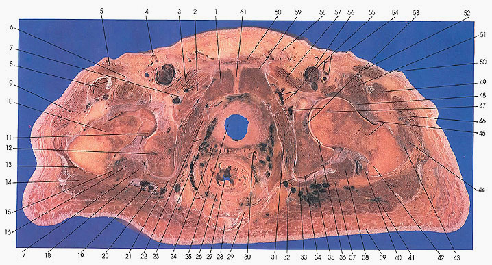
Anatomy Atlases Atlas Of Human Anatomy In Cross Section 6 Pelvis Perineum Hip And Upper Thigh from www.anatomyatlases.org Instant anatomy is a specialised web site for you to learn all about human anatomy of the body with diagrams, podcasts and revision questions Muscles of the lower limb; Case contributed by dr roberto schubert. This webpage presents the anatomical structures found on ankle mri. A cross section of the thigh vectorized and adapted from figure 432 from the 1918 grays anatomy. Instant anatomy is a specialised web site for you to learn all about human anatomy of the body with diagrams podcasts and revision questions. It consists of three muscle compartments (anterior, posterior, medial) which create movement by acting on the femur bone. Lower limb sectional anatomy abbas a.
• the composition and construction features of the four major joints (sacroiliac joint, thigh, knee and talocrural joint);
Each compartment has a distinct innervation and function. Muscle the lies over the frontal bone. The book is designed to help novices acquire pattern recognition skills to resolve. Harry benjamin laing, mrcs, ortho m8, frcs(tr and orth) tutorials A cross section of the thigh vectorized and adapted from figure 432 from the 1918 grays anatomy. Muscles of the lower limb; 1 article features images from this case. This mri knee sagittal cross sectional anatomy tool is absolutely free to use. Like the biceps brachii in the arm, the biceps femoris muscle has two heads. Sets of questions include anatomical images in the axial (transverse) plane. Anatomical structures of the lower limb (hip, thigh, knee, leg, ankle and foot) and specific regions (compartment of the lower limb) are visible on dynamic labeled images. The three layers of gluteal muscles, gluteus maximus, gluteus medius, gluteus minimus.like the forearm, the upper leg, or thigh, has a dense arrangement of many muscles.on the anterior side, the most prominent of the muscles are the sartorius muscle and the four muscles. Anatomy of the thigh and leg the thigh is best described in terms of compartmental anatomy, and is composed of anterior, posterior, and medial (adductor) compartments.
The muscles of the lower limb are numerous and complex. Upper thigh cross sectional anatomy : This mri wrist coronal cross sectional anatomy tool is absolutely free to use. The hamstring portion of the adductor magnus has a similar action to these muscles, but is located in the medial thigh. Stanford bone tumor ddx | iss/ssr msk lectures | search ocad cases | stanford virtual readouts stanford msk mri atlas has served over 1,000,000 pages to users in over 100 countries.
Mri Pelvis Anatomy Free Male Pelvis Axial Anatomy from mrimaster.com Radiologists perform ankle imaging to assess. These questions correlate anatomical structures with clinical problems. Muscles adapted for loaded versus unloaded actions. Shawka medical student 1st stage 2. Their origins and insertions are difficult to remember, and they are best considered as parts of general functional groups. The rectus femoris is located in the center of the thigh, while the vastus medialis is in the middle of the said body part. This mri hip joint axial cross sectional anatomy tool is absolutely free to use. Cross sectional anatomy of distal lower leg.
Anatomy of the thigh and leg the thigh is best described in terms of compartmental anatomy, and is composed of anterior, posterior, and medial (adductor) compartments.
Iliopsoas psoas major psoas minor iliacus buttocks gluteal r. Anatomy of the thigh and leg the thigh is best described in terms of compartmental anatomy, and is composed of anterior, posterior, and medial (adductor) compartments. Tendons are cords made of tough tissue, and they work as special connector pieces between bone and muscle. Anatomical structures of the lower limb (hip, thigh, knee, leg, ankle and foot) and specific regions (compartment of the lower limb) are visible on dynamic labeled images. The muscles of the lower limb are numerous and complex. • the groups, delamination, and position relation of muscles of lower limb; This webpage presents the anatomical structures found on ankle mri. This mri knee sagittal cross sectional anatomy tool is absolutely free to use. Serial cross sections variant image id: The muscles located within the posterior compartment of the thigh are the biceps femoris, semitendinosus and semimembranosus. Each compartment has a distinct innervation and function. Their origins and insertions are difficult to remember, and they are best considered as parts of general functional groups. 1 article features images from this case.
Meanwhile, the vastus lateralis is on the side of the thigh, while the vastus intermedius is hidden below the rectus femoris(5) upper thigh anatomy. Anatomy of the thigh and leg the thigh is best described in terms of compartmental anatomy, and is composed of anterior, posterior, and medial (adductor) compartments.


0 Komentar